See for yourself how the SEM works
 High Magnification High Magnification
Bulk sample analysis using the SEM is our standard service at
no extra charge. Our UKAS accredited and experienced laboratory
team analyse samples using an Scanning Electron Microscope (SEM)
which has a higher magnification of 300,000x in comparison to the
industry standard Polarised Light Microscope (PLM) which is restricted
to a magnification of no more than 1000x.
The industry standard PLM is limited as the wavelength of light,
the method used for magnification by PLM, cannot show things that
are smaller than its own wavelength.
 Focus Focus
As some samples do not have flat surfaces, under magnification
the bumps show as mountain landscapes. A light microscopy would
only be able to focus only on the peaks, or the valleys, or points
in between. The SEM can have all of these features in focus at
the same time, enabling us to see and understand the true shape
we are looking at.
 Clarity Clarity
The images produced by the SEM are easy to understand as objects
the output is a “sensible looking” picture, not a indistinguishable
image that requires years of practice to understand its real-world
significance.
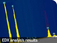 EDX analysis results EDX analysis results
When samples are viewed from an SEM, the x-rays generated by the
sample are analysed using an Energy Dispersive X-ray (EDX) component
which illustrates a qualitative elemental analysis on samples being
analysed, providing you with accurate results.
|